Overview

This page provides an introduction to atrial rhythms and links to our EKG interpretation courses and drills.
Atrial rhythms originate in the atria rather than in the SA node. The P wave will be positive, but its shape can be different from a normal sinus rhythm because the electrical impulse follows a different path to the AV (atrioventricular) node. These EKG differences are covered on our atrial rhythms training module as well as in practice strips which are available via a link in the right column. Atrial rhythms are classified as:
- Atrial Fibrillation (afib)
- Atrial Flutter
- Multifocal Atrial Tachycardia
- Premature Atrial Complex
- Supraventricular Tachycardia
- Wandering Atrial Pacemaker
- Wolff-Parkinson-White Syndrome
Atrial Rhythm Categories
Atrial Fibrillation

Irritable sites in the atria fire very rapidly, between 400-600 bpm. This very rapid pacemaking causes the atria to quiver. The ventricles beat at a slower rate due to the AV node's blocking some of the atrial impulses.
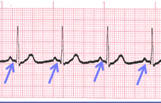
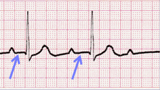
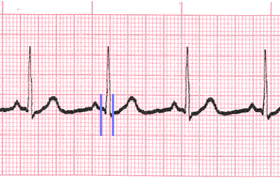
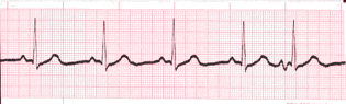
Atrial Flutter

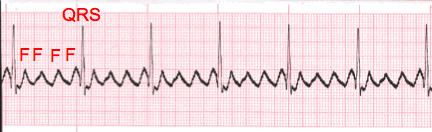
There are two types of atrial flutter. Type I (also called classical or typical) has a rate of 250-350 bpm. Type II (also called non-typical) are faster, ranging from 350-450 bpm. EKG tracings will show tightly spaced waves or saw-tooth waveforms (F-waves).
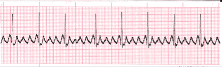
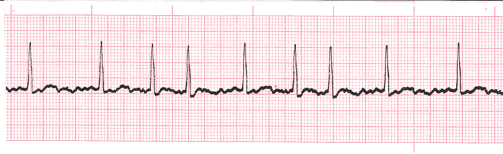
Multifocal Atrial Tachycardia

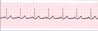
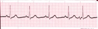
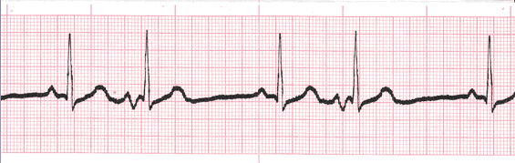
When multifocal atrial tachycardia occurs, multiple (non-SA) sites are firing impulses. The P waves will vary in shape and at least three different shapes can be observed. The PR Interval varies. Ventricular rhythm is irregular.
Premature Atrial Complex

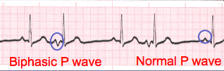
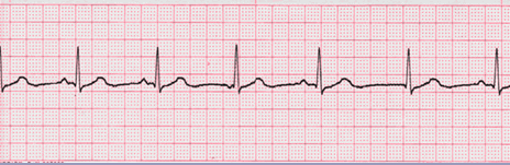
This occurs when an ectopic site within the atria fires an impulse before the next impulse from the SA node. If the ectopic site is near the SA node, the P wave will likely have a shape similar to a sinus rhythm. But this P wave will occur earlier than expected.
Supraventricular Tachycardia

This term covers three types of tachycardia that originate in the atria, AV junction or SA node.
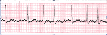
Wandering Atrial Pacemaker

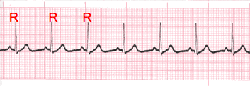
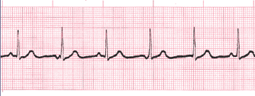
Wandering atrial pacemaker is an irregular rhythm. In is similar to multifocal atrial tachycardia but the heart rate is under 100 bpm. P waves are present but will vary in shape.
Wolff-Parkinson-White Syndrome

This occurs when the impulse travels between the atria and ventricles via an abnormal path, called the bundle of Kent. The impulse, not being delayed by the AV node, can cause the ventricles to contract prematurely. EKG characteristics include a shorter PR Interval, longer QRS complex and a delta wave.