using System.Threading;

Sinus Rhythms

What is a Sinus Rhythm?

sinus rhythm heart illustration

This page provides an introduction to sinus rhythms and links to our EKG interpretation courses and drills.

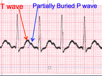
A normal sinus rhythm refers to both a normal heart rate and rhythm. Normal heart rates are from 60 to 100 beats per minute. The shape of the electrocardiogram (EKG) tracing will exhibit certain key attributes to be considered normal, as discussed below. With normal sinus rhythms, the heart beat's electrical impulse originates in the sinoatrial node (SA). The P waves are upright and appear before each QRS and have the same shape. The intervals between the P waves are regular, although some variations can occur with respiration. Sinus rhythms are classified as:

  • Normal Sinus Rhythm
  • Sinoatrial Block
  • Sinus Pause
  • Sinus Arrhythmia
  • Sinus Bradycardia
  • Sinus Tachycardia

Sinus Rhythm Categories
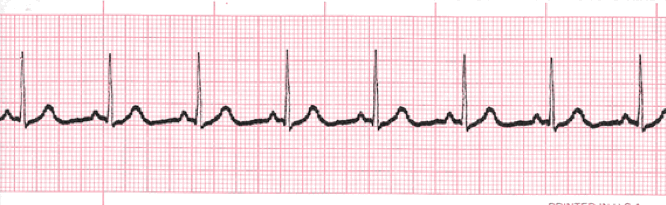
Normal Sinus Rhythm

normal sinus rhythm EKG tracing

This is the normal sinus rhythm of the heart. The electrical impulse originates within the SA node and travels through the atria to the AV node. After a brief delay, the impulse travels down the bundle branches, thought the Purkinje fibers to the ventricles.
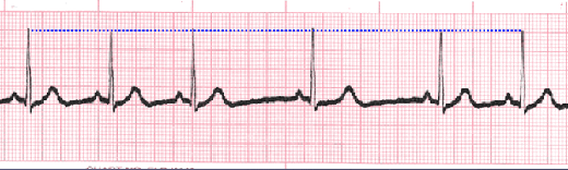
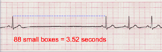
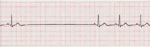
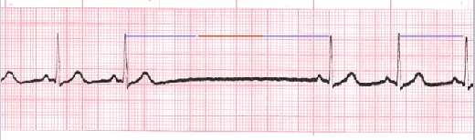
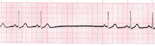
Sinus Pause

sinus pause EKG tracing

Sinus pause includes sinus arrest and sinus exit block. Sinus arrest is caused by failure of the SA node to create an impulse. An interruption in R-R regularity can be observed. With sinus exit block the SA node generates an impulse but it is blocked before being transmitted through the atria. R-R regularity continues with the beats that follow the missed beat. Depressed ST segments can also be observed.
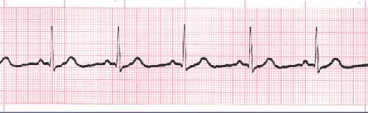
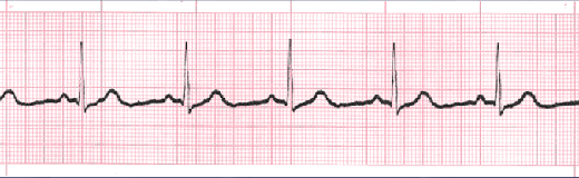
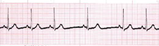
Sinus Arrhythmia

Sinus Arrhythmia EKG tracing

Sinus arrhythmia looks normal except for slight irregularities. A frequent cause of sinus arrhythmia can be rhythm variations caused by respiration.
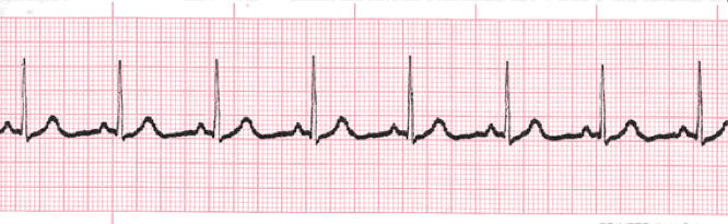
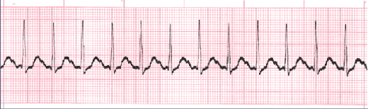
Sinus Bradycardia

Sinus Bradycardia EKG tracing

Sinus bradycardia is a sinus rhythm with a rate of 40-60 bpm.

Sinus Tachycardia

Sinus Tachycardia EKG tracing

Sinus tachycardia is a normal sinus rhythm, but with a heart rate over 100 bpm. It is a normal response to exercise, excitement and some illnesses.

Training Resources

Sinus Rhythms Training

A good starting point for learning is our sinus rhythms module. This module focuses on the morphologic features and qualifying criteria of sinus rhythms. A brief review of cardiac rhythm analysis in also included.

Sinus Rhythms Lessons


EKG Rhythm Tests

Hundreds heart rhythms in this practice test. Test can be tailored for specific learning needs.

EKG Rhythm Tests


Heart Rhythm Quiz

www.PracticalClinicalSkills.com is an excellent source of arrhythmia information and drills.

Sinus Arrhythmia Quiz


External References

Asystole
Wikipedia
Free Dictionary

Click To Begin Sinus Rhythms Training Module


Overview

Sinus Rhythms

Thomas E. O'Brien
AS CCT CRAT RMA

Learning Objectives

At the conclusion of this training module the reader will be able to:

  • Recall and apply the 5-steps of heart rhythm interpretation
  • Recognize the difference between regular and irregular rhythms
  • Recall the normal range for PR interval and QRS complex
  • Recognize the features and qualifying criteria for the following complexes and rhythms:
    • Normal Sinus Rhythm
    • Sinus Bradycardia
    • Sinus Tachycardia
    • Sinus Dysrhythmia (Arrhythmia)
    • Sinus Arrest
    • Sinus Exit Block

Interpretation

Introduction

  • The previous slides presented the five-steps of rhythm analysis. These five steps must be followed regardless of how simple of complex the tracing is you are reviewing.
  • The information gathered in these steps are telling a story.
  • The title of that story is the interpretation.

Sinus Rhythm Types

The dysrhythmias in this category occur as a result of influences on the Sinoatrial (SA) node. Rhythms in this category will share similarities in a normal appearing P wave, the PR interval will measure in the “normal range” of 0.12 – 0.20 second, and the QRS typically will measure in the “normal range” of 0.06 – 0.10 second. For the most part, dysrhythmias in this category either effect the rate, rhythm regularity or both within a particular tracing. We will be discussing the following complexes and rhythms:

  • Normal Sinus Rhythm
  • Sinus Bradycardia
  • Sinus Tachycardia
  • Sinus Dysrhythmia (Arrhythmia)
  • Sinus Arrest
  • Sinus Exit Block

Normal Sinus Rhythm

Introduction

normal sinus rhythm ekg
  • Also known as Sinus Rhythm is the only rhythm when each of the five steps of rhythm analysis are “normal”.
  • All other rhythms you will analyze will have at least one of the 5-steps presenting an abnormality.
  • This rhythm will be regular, in a heart rate range between 60 – 100 bpm, P waves are upright and uniform in appearance (in Lead II), with a P wave for each QRS complex, the PR interval will measure in the normal range of 0.12 – 0.20 second (and measure the same each time), and the QRS complex morphology will be similar beat-to-beat and measure between 0.06 – 0.10 second (and measure the same each time).

Practice Strip

normal sinus rhythm ekg
  • Analyze this tracing using the five steps of rhythm analysis.
  • Compare your answers with the answers on the next slide.

Answers

normal sinus rhythm ekg
  • Rhythm: Regular
  • Rate: 75 (20 small boxes)
  • P wave: Upright & uniform
  • PR interval: 0.16 second
  • QRS: 0.08 second
  • Normal Sinus Rhythm

Sinus Bradycardia

Description

sinus bradycardia tracing
  • Rhythms are often named according to the origin of the electrical activity in the heart or the structure where the problem is occurring.
  • Sinus Rhythms are aptly named due to the locus of stimulation being the SA (sinoatrial) node.
  • With Sinus Bradycardia the locus of stimulation is the same as normal sinus rhythm, just now the rate will be less than 60 bpm. Recall that “brady” means slow.
  • The only difference between sinus bradycardia and normal sinus rhythm is the rate. All other steps in the rhythm analysis are “normal”.

Practice Strip

Sinus Bradycardia - Practice Strip

sinus bradycardia tracing
  • Analyze this tracing using the five steps of rhythm analysis.
  • Compare your answers with the answers on the next slide.

Answers

Practice Strip - Answers

sinus bradycardia tracing
  • Rhythm: Regular
  • Rate: 56 (27 small boxes)
  • P wave: Upright and Uniform
  • PR interval: 0.18 sec
  • QRS: 0.10 sec
  • Interpretation: Sinus Bradycardia

Sinus Tachycardia

Description

sinus tachycardia tracing
  • Sinus Tachycardia occurs when the rate of electrical impulse formation occurs at a rate exceeding 100 bpm.
  • This can occur for a number of different reasons i.e. diet, stress, illness, response to physical exertion etc.
  • The only difference between Normal Sinus Rhythm and Sinus Tachycardia is the rate exceeds 100 bpm. All other steps of rhythm analysis will be “normal.
  • An additional challenge that will present as rhythm rates accelerate is that the cardiac complexes will come closer together. This can result in the P wave becoming partially or completely buried within the T wave of the previous cardiac complex.
  • The result of a partially buried P waves means you are unable to establish the beginning of atrial depolarization. Meaning you will be unable to measure and report the PR interval. The only way it will be possible is if the physician instructs you to increase the machine printing speed (remember interval times will double at 50 mm/sec).

Practice Strip

Sinus Tachycardia - Practice Strip

sinus tachycardia tracing
  • Analyze this tracing using the five steps of rhythm analysis.
  • Compare your answers with the answers on the next slide.

Answers

Practice Strip - Answers

sinus tachycardia tracing
  • Rhythm: Regular
  • Rate: 125
  • P wave: upright, partially buried
  • PR interval: unable to determine
  • QRS: 0.08 sec
  • Interpretation: Sinus Tachycardia

Sinus Dysrhythmia (Arrhythmia)

Description

sinus arrhythmia tracing
  • Sinus Dysrhythmia often occurs as a normal variant. It is commonly seen in young healthy people and athletes. It is frequently related to breathing and pressure on the vagus nerve. As the patient inhales and the lungs expand, pressure is applied to the vagus nerve which causes a parasympathetic response and a decrease in heart rate.
  • Sinus Dysrhythmias may also occur as a result of medication effects or diseases such as multiple sclerosis.
  • This dysrhythmia often requires no treatment, but may require medication or therapy such as a pacemaker to regulate the heart rate if the ventricular response becomes too slow.
  • Sinus Dysrhythmia closely resembles Normal Sinus Rhythm with the only distinction being the intervals from one cardiac complex to the next are changing as influenced by the patients respiratory pattern.
  • Note the changing R to R Intervals.

Practice Strip

Sinus Tachycardia - Practice Strip

sinus dysrhythmia tracing
  • Analyze this tracing using the five steps of rhythm analysis.
  • Compare your answers with the answers on the next slide.

Answers

Practice Strip - Answers

sinus dysrhythmia tracing
  • Rhythm: Irregular
  • Rate: 60 bpm
  • P wave: upright and uniform
  • PR interval: 0.16 sec
  • QRS: 0.10 sec
  • Interpretation: Sinus Dysrhythmia

Sinus Arrest

Description

  • Sinus Arrest occurs when there is a sudden absence of electrical activity initiated by the SA node. This results in a pause in the electrical activity seen on the tracing. Remember, no electrical activity = no depolarization and contraction. Hence, a drop in blood pressure.
  • The longer the pause, the further the blood pressure will drop.
  • A pause of six-seconds is considered a medical emergency and emergency procedures must be initiated.
  • The rhythm typically will demonstrate constant R to R intervals prior to and following the pause. This pause results in the rhythm tracing presenting as irregular.

Analysis

sinus arrest ekg tracing
  • It is important to measure the duration of the pause and report this information along with the frequency of the pauses if there are more than one.
  • The pause is measured by placing your caliper tips on the last R wave just prior to the pause and the R wave immediately following the pause. Count the number of small boxes and multiply by 0.04 second. This will be reported as part of the interpretation.

Practice Strip

Sinus Arrest - Practice Strip

sinus arrest ekg tracing
  • Analyze this tracing using the five steps of rhythm analysis.
  • Compare your answers with the answers on the next slide.

Answers

Practice Strip - Answers

sinus arrest ekg tracing
  • Rhythm: Irregular
  • Rate: 40 bpm
  • P wave: upright and uniform
  • PR interval: 0.16 sec
  • QRS: 0.10 sec
  • Interpretation: Sinus Arrest, 3.52 second pause

Sinus Exit Block

Description

sinus exit block ekg tracing
  • Sinus Exit Block looks very much the same as Sinus Arrest with one important distinction.
  • The duration of the pause with Sinus Exit Block is in a direct multiple of the R to R interval of the underlying rhythm. Sinus Arrest does not have this specific feature.

Practice Strip

Sinus Dysrhythmia - Practice Strip

sinus exit block tracing
  • Analyze this tracing using the five steps of rhythm analysis.
  • Compare your answers with the answers on the next slide.

Answers

Practice Strip - Answers

sinus exit block tracing
  • Rhythm: Irregular
  • Rate: 40 bpm
  • P wave: upright and uniform
  • PR interval: 0.16 sec
  • QRS: 0.10 sec
  • Interpretation: Sinus Exit Block


EKG Self Test

EKG Rhythms Self Test

If you would like to test the knowledge and skills that you have learned in this module, use our EKG Rhythms Self Test. You can choose to focus this self-test on any or all of the following:

  • Sinus Mechanisms
  • Atrial Rhythms
  • Junctional Rhythms
  • Ventricular Rhythms
  • Atrioventricular Blocks
  • Pacemaker Rhythms

EKG Rhythm Tests


Authors and Reviewers


Sources

Authors and Reviewers


Sources


? v:1 | cc: | tar: False | onAr:0 | onPs:2 | tLb:1 | tLbJs:0
| pv: 1 uStat: False | db:0 | shouldInvoke:False | pu:False | em: | refreshTime: 1/1/0001 12:00:00 AM || now: 4/24/2024 11:30:43 PM | firstPage: True
| statusStg: | cDbLookup# 0


An error has occurred. Please reload the page or visit our other website, Practical Clinical Skills. Reload 🗙