ECG Axis Determination EKG
Your online EKG class
ECG Axis Determination
Thomas E. O'Brien
AS CCT CRAT RMA
Learning Objectives
Upon completion of the accompanying narrative and practice the student will be able to:
- Recall which major coronary artery supports each region of the heart
- Associate the views of a 12-Lead ECG with specific surfaces of the heart
- Determine the presence of cardiac axis deviation
- Understand Einthoven's Triangle
12-Lead ECG
- People may choose to analyze ECG’s in a number of different ways. The sequence doesn’t necessarily matter as long as you gather and report the proper information each time. I read from left to right as much as possible (in anatomic groupings).
- If your protocols are different, always follow them.
- The following slides will review the leads, surfaces, and associated coronary arteries which commonly supply that portion of the heart.
Lessons
Lesson #1: Views 322
II, III, and aVF
- Inferior wall of left ventricle
- Right Coronary Artery
- Marginal branch
V1 and V2
- Septum
- Left Coronary
- Septal branch
V3 & V4
- Anterior
- Left Coronary
- Anterior Descending and Diagonal arteries
I, aVL, V5 & V6
- Lateral
- Left Coronary
- Circumflex & Obtuse marginal
Labeled Views

Lesson #2: Quick Check Views 1
Question 1
Name the inferior view leads.
Answer
II, III & aVF
Question 2
Name the septal view leads.
Answer
V1 & V2
Lesson #3: Quick Check Views 2
Question 1
Name the anterior view leads.
Answer
V3 & V4
Question 2
Name the lateral view leads.
Answer
I, aVL, V5 & V6
Lesson #4: Axis & Deviation
Description
- Axis is the general flow of electricity as it passes through the heart from the SA node all the way to the Purkinje fibers.
- It is typically obtained from the limb or frontal plane leads.
- The axis of the heart can change for a number of different reasons from birth defects which affect the physical location of the heart i.e. Dextrocardia, to obesity to pregnancy among others.
- If you can envision a circle with a centered vertical and horizontal line superimposed over the chest, you would note that the normal heart is located in the lower left quadrant of this circle.
- You will be provided an example of this image in a few slides.
Electrical Axis Determination
Axis determination can be determined a number of different ways. From complex to simple. The following steps are the easiest I know to obtain this important information.
Electrical Axis Determination
Imagine a large circle, divided into four equal quadrants superimposed over the patient’s chest.

Lesson #5: Axis Determination
Part I
- The direction of the ventricular depolarization (QRS complex) is analyzed.
- Refer to lead I and aVF each time
- If the majority of the QRS complex is positively deflected in both views, this is referred to as “normal axis”.


Part II
If lead I is positive and aVF is negative, this is ‘left axis deviation”


Part III
If lead I is negative and aVF is positive, this is “right axis deviation”.
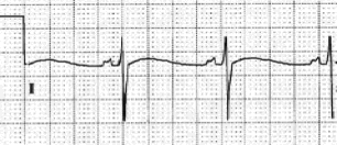
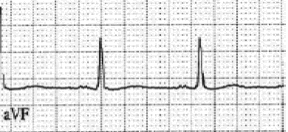
Part IV
If lead I is negative and aVF is negative, this is extreme right axis deviation aka indeterminate.
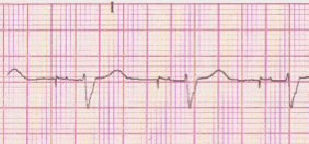
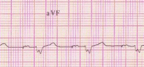
Lesson #6: Quick Check Axis 1
Question
Identify the electrical axis
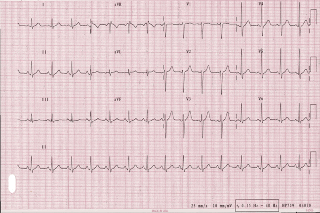
Answer
Normal Axis – Lead I up, aVF up
Question
Identify the electrical axis
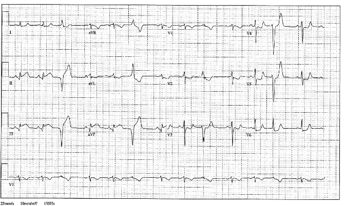
Answer
Despite the presence of premature ventricular complexes, when analyzing the ventricular depolarization of the underlying rhythm you will note Normal Axis – Lead I up, aVF up
Question
Identify the electrical axis

Answer
Normal Axis – Lead I up, aVF up
Lesson #7: Quick Check Axis 2
Question
Identify the electrical axis
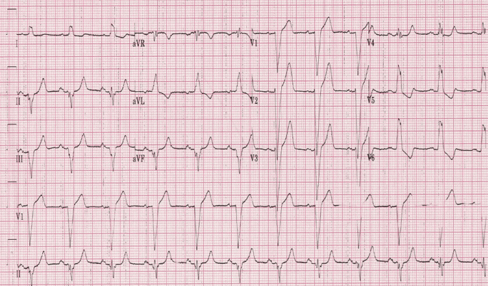
Answer
Left Axis – Lead I up, aVF down
Question
Identify the electrical axis
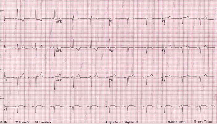
Answer
Left Axis – Lead I up, aVF down
Question
Identify the electrical axis
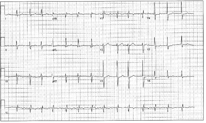
Answer
Left Axis – Lead I up, aVF down
Lesson #8: Quick Check Axis 3
Question
Identify the electrical axis
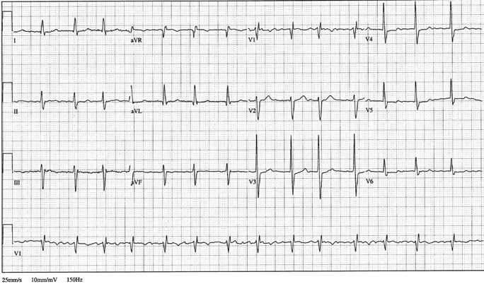
Answer
Left Axis – Lead I up, aVF down
Question
Identify the electrical axis
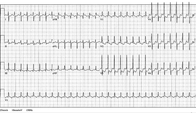
Answer
Right Axis – Lead I down, aVF up
Question
Identify the electrical axis
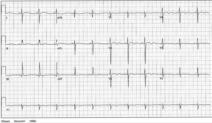
Answer
Right Axis – Lead I down, aVF up
Lesson #9: Quick Check Axis 4
Question
Identify the electrical axis
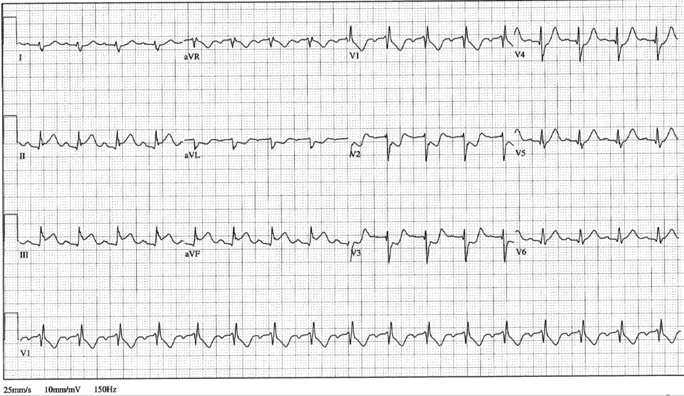
Answer
Right Axis – Lead I down, aVF up
Question
Identify the electrical axis
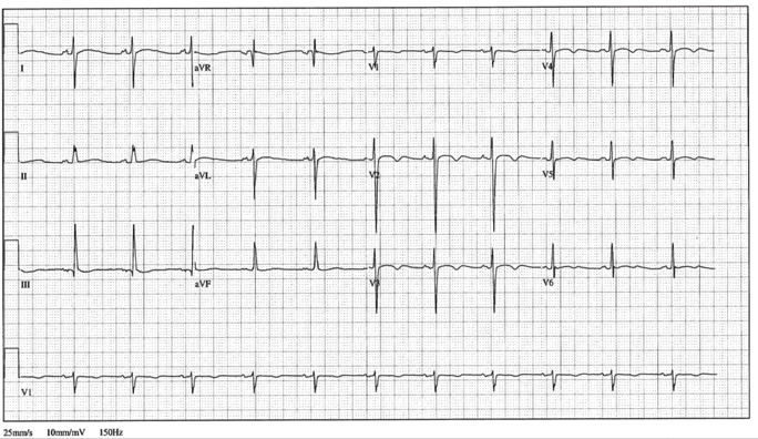
Answer
Right Axis – Lead I down, aVF up
Question
Identify the electrical axis
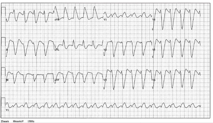
Answer
Extreme Right Axis – Lead I down, aVF down
Lesson #10: Quick Check Axis 5
Question
Identify the electrical axis
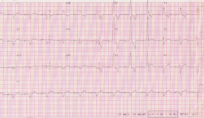
Answer
Extreme Right Axis – Lead I down, aVF down
Question
Identify the electrical axis
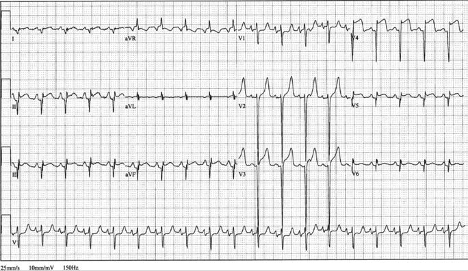
Answer
Right Axis – Lead I down, aVF up
Return to Main Lessons Page
Authors and Reviewers
- EKG heart rhythm modules: Thomas O'Brien.
- EKG monitor simulation developer: Steve Collmann
-
12 Lead Course: Dr. Michael Mazzini, MD.
- Spanish language EKG: Breena R. Taira, MD, MPH
- Medical review: Dr. Jonathan Keroes, MD
- Medical review: Dr. Pedro Azevedo, MD, Cardiology
- Last Update: 11/8/2021
Sources
-
Electrocardiography for Healthcare Professionals, 5th Edition
Kathryn Booth and Thomas O'Brien
ISBN10: 1260064778, ISBN13: 9781260064773
McGraw Hill, 2019 -
Rapid Interpretation of EKG's, Sixth Edition
Dale Dublin
Cover Publishing Company -
12 Lead EKG for Nurses: Simple Steps to Interpret Rhythms, Arrhythmias, Blocks, Hypertrophy, Infarcts, & Cardiac Drugs
Aaron Reed
Create Space Independent Publishing -
Heart Sounds and Murmurs: A Practical Guide with Audio CD-ROM 3rd Edition
Elsevier-Health Sciences Division
Barbara A. Erickson, PhD, RN, CCRN -
The Virtual Cardiac Patient: A Multimedia Guide to Heart Sounds, Murmurs, EKG
Jonathan Keroes, David Lieberman
Publisher: Lippincott Williams & Wilkin)
ISBN-10: 0781784425; ISBN-13: 978-0781784429 - Project Semilla, UCLA Emergency Medicine, EKG Training Breena R. Taira, MD, MPH