Spirometry EKG
Your online EKG class
Spirometry Definition
Quite simply, spirometry measures two basic elements:
1) The amount of usable air in the lungs (vital capacity)
2) How fast the patient can blow the air out
Spirometry is a component of pulmonary function testing. While a complete pulmonary function test takes place in a dedicated lab, spirometry can be performed at the bedside or in an office.
PowerPoint Slides
We have created a Spirometry Presentation to introduce the principles of spirometry. This presentation also provides a introduction to interpretation of the values obtained. The author has established these objectives for the learner:
- Define the basic lung volumes and capacities
- Describe how the test is performed and the values obtained
- Describe basic qualities assurance measures to ensure the accuracy of the results
- State the significance of the data, especially for COPD and asthma
- Given a set of spirometry data, provide a basic interpretation
Spirometry Interpretation - Data Review
What Data Tells You
Once the testing is complete, review the data obtained and ask questions such as:
- Is it a good test, that is, are you satisfied that the subject put forth the best effort possible?
- To determine this, look at the data: Do you have two sets of data that are close in value; do the flow/volume loops overlap? Also, look at the PEFR.
- Are all the values within normal limits (80% - 120% of what was predicted for the subject)?
- If the values are not all within normal limits, is the abnormality more indicative of a restrictive disorder or an obstructive disorder (or possibly both)? If the FEV1, FEV1/FVC, and/or FEF25%-75% is low, the abnormality is most likely obstructive. If the flows are normal but the FVC is low, the abnormality is most likely restrictive (however, this needs to be confirmed by measuring the TLC in a PFT lab).
Spirometry Normal Values
Normal values for each data point obtained during spirometry is derived from a predictive equation and is patient specific. The predictive equations require the patient’s gender, height, weight, and age. Some predictive equations also include race. Fortunately, nearly all modern spirometers automatically calculate the patient’s predicted values. The data point is generally considered normal if it falls within 80% to 120% of what has been predicted.

Spirometry Terms & Concepts
- Total Lung Capacity (TLC). The maximum amount of air the lung can hold.
- Vital Capacity (VC). The maximum amount of air that can be exhaled following a maximum inhalation.
- Forced Vital Capacity (FVC). The vital capacity obtained when the subject is blowing out as hard and fast as possible.
- Residual Volume (RV). The amount of air remaining in the lungs following a maximum exhalation.
- Functional Residual Capacity (FRC). The amount of air remaining in the lungs following a normal exhalation.
- Volume. Recall that spirometry measures the amount of usable air in the lungs. This is the vital capacity. And because the subject is asked to blow out as hard and fast as possible, the value obtained is the forced vital capacity.
- Residual Volume. Note that spirometry cannot measure residual volume, functional residual capacity, or total lung capacity since residual volume cannot be exhaled. These values need to be obtained by other means in a pulmonary function lab.
- Flow. Recall that part of spirometry is determining how fast the subject is able to exhale (or blow out) their vital capacity. The primary reason this is measured is because in certain diseases such as COPD and asthma, the patient’s ability to exhale is compromised.
There are generally three or four measures of a subject’s ability to exhale hard and fast.
- Forced Expired Volume in the first second (FEV1). This is a measure of how much of the FVC the subject exhaled in the first second. A normal person will exhale around 80% of their FVC in the first second.
- FEV1/FVC. This is the percent of the FVC that person exhaled in the first second (see above). Both measures are important to look at together because if the FVC is low, the FEV1 will also be low (as in restrictive diseases such as idiopathic pulmonary fibrosis; however in these diseases, the person’s ability to exhale is not usually compromised).
Other Flow Measures
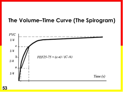
- Forced Expiratory Flow between 25% and 75% (FEF25-75). This is the average flowrate calculated as the subject is exhaling between 25% and 75% of his/her FVC. Think about it this way: As the subject forcibly exhales the FVC, the rate at which the flow is leaving the lungs is not constant, but rather changes over the length of exhalation.
- Peak Expiratory Flow (PEF or sometimes PEFR). The highest flowrate the subject attains during exhalation. It usually occurs at the beginning of exhalation. Often used as an indication of effort.
Sample Graphs


Lessons
Lesson #1: Introduction
Introduction

Disclaimer
This presentation is intended to be a basic introduction to the principles of spirometry. It will also touch briefly on the interpretation of the values obtained.
The presentation is not intended to be definitive or a substitute for a formal course in pulmonary function testing. For more detail, the learner is directed to several good textbooks and textbook chapters. There are also a number of short videos on youtube.com that serve to demonstrate spirometry.
Objectives
Upon successful completion of this presentation, the learner will be able to:
- Define the basic lung volumes and capacities
- Describe how the test is performed and the values obtained
- Describe basic qualities assurance measures to ensure the accuracy of the results
- State the significance of the data, especially for COPD and asthma
- Given a set of spirometry data, provide a basic interpretation
Using This Presentation

Lesson #2: Spirometry and PFT
What is Spirometry
Quite simply, spirometry measures two basic elements:
- The amount of usable air in the lungs (vital capacity)
- How fast the patient can blow the air out
Pulmonary Function Testing
- Spirometry is a component of pulmonary function testing.
- Complete pulmonary function testing must be performed in a dedicated lab.
- Spirometry, as a stand alone procedure, can be done at the bedside or in the office setting.
Lesson #3: Reasons to Perform Spirometry
Part 1
- Aid in the diagnosis and staging of COPD
- Aid in the diagnosis of asthma
- Aid in determining appropriate treatment for patients with shortness of breath
Part 2
- Aid in tracking lung function over time (either improvement or deterioration) in patients with certain chronic diseases (e.g. sarcoidosis, idiopathic pulmonary fibrosis, neuromuscular, etc.)
- Screening patients who are asymptomatic but at risk for developing lung disease (e.g. smokers)
Lesson #4: The Lungs
Volumes & Capacities
The lung can be divided into eight volumes and capacities:

Lesson #5: Definitions & Nornal Values
Normal Values
- Normal values for each data point obtained during spirometry is derived from a predictive equation and is patient specific. The predictive equations require the patient’s gender, height, weight, and age. Some predictive equations also include race.
- Fortunately, nearly all modern spirometers automatically calculate the patient’s predicted values.
- The data point is generally considered normal if it falls within 80% to 120% of what has been predicted.
Definitions
| Total Lung Capacity (TLC) | The maximum amount of air the lung can hold. |
| Vital Capacity (VC) | The maximum amount of air that can be exhaled following a maximum inhalation. |
| Forced Vital Capacity (FVC) | The vital capacity obtained when the subject is blowing out as hard and fast as possible. |
| Residual Volume (RV) | The amount of air remaining in the lungs following a maximum exhalation. |
| Functional Residual Capacity (FRC) | The amount of air remaining in the lungs following a normal exhalation. |
Lesson #6: Measurements
Volume
- Recall that spirometry measures the amount of usable air in the lungs. This is the vital capacity. And because the subject is asked to blow out as hard and fast as possible, the value obtained is the forced vital capacity.
- Note that spirometry cannot measure residual volume, functional residual capacity, or total lung capacity since residual volume cannot be exhaled. These values need to be obtained by other means in a pulmonary function lab.
Flow
- Recall that part of spirometry is determining how fast the subject is able to exhale (or blow out) their vital capacity. The primary reason this is measured is because in certain diseases such as COPD and asthma, the patient’s ability to exhale is compromised.
- There are generally three or four measures of a subject’s ability to exhale hard and fast.
Lesson #7: Measures
Measures
- Forced Expired Volume in the first second (FEV1):
This is a measure of how much of the FVC the subject exhaled in the first second. A normal person will exhale around 80% of their FVC in the first second. - FEV1/FVC:
This is the percent of the FVC that person exhaled in the first second (see above). Both measures are important to look at together because if the FVC is low, the FEV1 will also be low (as in restrictive diseases such as idiopathic pulmonary fibrosis; however in these diseases, the person’s ability to exhale is not usually compromised).
Other Flow Measures
- While the FEV1 and the FEV1/FVC are the most useful flow measures, others are available and often obtained.
- Forced Expiratory Flow between 25% and 75% (FEF25-75): This is the average flowrate calculated as the subject is exhaling between 25% and 75% of his/her FVC. Think about it this way: As the subject forcibly exhales the FVC, the rate at which the flow is leaving the lungs is not constant, but rather changes over the length of exhalation.
- Peak Expiratory Flow (PEF or sometimes PEFR): The highest flowrate the subject attains during exhalation. It usually occurs at the beginning of exhalation. Often used as an indication of effort.
Lesson #8: Performing a Test 1
How a Test is Performed
- Prepare patient and equipment
- Test Steps:
- patient inhales to total lung capacity
- blows out hard and fast to residual volume
- inhales to total lung capacity
- Repeat test
- Record data
Tips
- Prepare equipment according to standards set by the American Thoracic Society or European Respiratory Society
- Gather the following data from the test subject:
- age
- height
- weight
- any relevant history and/or purpose of the test
- Ensure a tight seal around the mouthpiece
Lesson #9: Performing a Test 2
Importance of Coaching
- Always remember that this is an effort-dependent test. This means that the person being tested must be both able and willing to blow out hard and fast to the best of their ability.
- This requires the test administrator (tech) carefully explain the nature of the test and what is expected. It is also helpful to assertively coach the testing subject during the performance of the test.
Quality Assurance
- The best way to ensure that the subject has performed the test to the best of his or her ability is for the subject to perform the test more than once – at least enough times to obtain repeatability, if possible.
- You can also look at the peak expiratory flowrate as a quasi measure of effort (i.e. if the PEFR is low but all other values are normal or near normal, the effort was probably poor).
Lesson #10: Results 1
Results

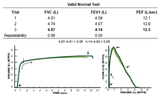
Flow/Volume Loops


Volume/Time Graph

Lesson #11: Results 2
Flow/Volume Loops


Normal Test

Normal Data Set

Multiple Efforts

Lesson #12: Interpretation Guidelines 1
Guidelines
- Normal for FVC, FEV1, FEF25%-75%, and PEFR is between 80% and 120% of what has been predicted for the patient.
- Often, the test is performed before and after the administration of a bronchodilator. Improvement is noted as in the table below:

Chart

Lesson #13: Interpretation Guidelines 2
What Data Tells You
- Once the testing is complete, look at the data obtained and ask yourself a succession of questions.
- 1) Is it a good test, that is, are you satisfied that the subject put forth the best effort possible?
- To determine this, look at the data: Do you have two sets of data that are close in value; do the flow/volume loops overlap? Also, look at the PEFR.
What Data Tells You (con't)
- 2) Are all the values within normal limits (80% - 120% of what was predicted for the subject)?
- 3) If the values are not all within normal limits, is the abnormality more indicative of a restrictive disorder or an obstructive disorder (or possibly both)? If the FEV1, FEV1/FVC, and/or FEF25%-75% is low, the abnormality is most likely obstructive. If the flows are normal but the FVC is low, the abnormality is most likely restrictive (however, this needs to be confirmed by measuring the TLC in a PFT lab).
Lesson #14: Degree of Severity
Severity
- 4) What is the degree of severity? This question can be answered for an obstructive problem. However, it cannot be answered for a restrictive problem. Degree of severity for restrictive is determined by the total lung capacity.
- FEV1 < 65-80% mild obstruction
- FEV1 < 50-65% moderate obstruction
- FEV1 < 50% severe obstruction
Reversibility
- 5) If a bronchodilator was administered and the test repeated, did the FEV1 or FVC improve (see interpretation guidelines above)?
Lesson #15: COPD
COPD Staging

COPD Sample

COPD (cont'd)
Possibly Mixed Obstructive & Restrictive

COPD (cont'd)
Possibly Mixed Obstructive & Restrictive
62 year-old female

Lesson #16: Try This One
Question
52 year-old male

Answer
- None of the five parameters is normal
- Because both the FEV1 and the FEV1/FVC are low, this is an obstructive pattern. Because the FVC is only 69% of predicted, there could also be a restrictive issue as well. However, this can only be confirmed by a decrease in the total lung capacity.
- Because the FEV1 < 50%, this is severe obstruction.
- No bronchodilator was administered.
Lesson #17: Recommendations
Recommendations
This has been a very basic introduction to spirometry. For more knowledge and practice, you are encouraged to do the following:
- Study relevant texts on pulmonary function testing
- Contact spirometer vendors for more information
- Watch relevant youtube videos
- Contact local pulmonary function labs in order to possibly observe technique and review data
Lesson #18: Short Quiz
Question #1
Which of the following cannot be obtained via spirometry?
B. FEV1
C. Peak flowrate
D. Residual volume
Question #2
During the performance of the forced vital capacity, the patient should do which of the following?
B. Exhale hard and fast to residual volume
C. Exhale slowly to functional residual capacity
D. Inhale fast then exhale fast
Question #3
The FVC is considered to be normal if it is which of the following?
B. At least 5 liters
C. At least 70% of predicted
D. At least 80% of predicted
Question #4
The severity of an obstructive disorder is determined on the basis of which of the following?
B. FEV1
C. FEF25%-75%
D. PEFR
Return to Main Lessons Page
Authors and Reviewers
- EKG heart rhythm modules: Thomas O'Brien.
- EKG monitor simulation developer: Steve Collmann
-
12 Lead Course: Dr. Michael Mazzini, MD.
- Spanish language EKG: Breena R. Taira, MD, MPH
- Medical review: Dr. Jonathan Keroes, MD
- Medical review: Dr. Pedro Azevedo, MD, Cardiology
- Last Update: 11/8/2021
Sources
-
Electrocardiography for Healthcare Professionals, 5th Edition
Kathryn Booth and Thomas O'Brien
ISBN10: 1260064778, ISBN13: 9781260064773
McGraw Hill, 2019 -
Rapid Interpretation of EKG's, Sixth Edition
Dale Dublin
Cover Publishing Company -
12 Lead EKG for Nurses: Simple Steps to Interpret Rhythms, Arrhythmias, Blocks, Hypertrophy, Infarcts, & Cardiac Drugs
Aaron Reed
Create Space Independent Publishing -
Heart Sounds and Murmurs: A Practical Guide with Audio CD-ROM 3rd Edition
Elsevier-Health Sciences Division
Barbara A. Erickson, PhD, RN, CCRN -
The Virtual Cardiac Patient: A Multimedia Guide to Heart Sounds, Murmurs, EKG
Jonathan Keroes, David Lieberman
Publisher: Lippincott Williams & Wilkin)
ISBN-10: 0781784425; ISBN-13: 978-0781784429 - Project Semilla, UCLA Emergency Medicine, EKG Training Breena R. Taira, MD, MPH