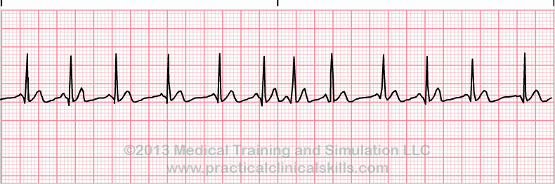
Multifocal Atrial Tachycardia EKG Interpretation with Rhythm Strip
EKG Features
Rate: Fast (> 100 bpm)Rhythm: Irregular
P Wave: Often changing shape and size from beat to beat (at least three differing forms)
PR Interval: Variable
QRS: Normal (0.06-0.10 sec)
A faster version of Wandering Atrial Pacemaker. Observe at least three different shaped P waves.
Multifocal atrial tachycardia is caused by electrical signals being sent from multiple (ectopic) locations in the atria rather than from the sinoatrial (SA) node. These multiple signals cause a rapid, inefficient heartbeat. This arrhythmia is more commonly found in patients over 50 years of age, particular in patients with lung disorders. Also see Wandering Atrial Pacemaker, a related abnormality.
Wikipedia
Multifocal Atrial Tachycardia EKG Interpretation Example
Return to EKG Reference Guide Index