Junctional Rhythms
Overview
This page provides an introduction to junctional rhythms and links to our EKG junctional rhythm interpretation training content.
The SA node is the normal origin of the electrical impulse for a heart beat. When the SA node cannot perform this role, the atrioventricular (AV) node may take over pacemaking. When this occurs, the EKG will likely have distinctive waveform features that reveal important aspects of these junctional rhythms.
Junctional rhythms include:
- Accelerated Junctional Rhythm
- Junctional Escape Rhythm
- Junctional Tachycardia
- Premature Junctional Complex
Junctional Rhythm Categories
Accelerated Junctional Rhythm

Accelerated junctional rhythm EKG occurs when the AV junction fires impulses at above 60 bpm. Rhythm will be very regular. The QRS complex is narrow (0.10 sec or less).
Junctional Escape Rhythm

Junctional escape beats originate in the AV junction and are late in timing. They often occur during sinus arrest or after premature atrial complexes. The QRS complex will be measured at 0.10 sec or less. Rhythm will be regular with a rate of 40-60 bpm.
Junctional Tachycardia

This abnormal rhythm originates in the bundle of His. It is observed as three or more premature junctional complexes (PJCs) appearing in a row. Heart rate will be over 100 bpm.
Premature Junctional Complex

Premature junctional complex (PJC) occurs when an irritable site within the AV node fires an impulse before the SA node. This impulse interrupts the sinus rhythm. The QRS complex will be narrow, usually measured at 0.10 sec or less.
Click To Begin Junctional Rhythms Module
Lessons
Lesson #1: Rhythm Analysis Method 314
Intro
The five steps of rhythm analysis will be followed when analyzing any rhythm strip. Analyze each tracing in the following order.
- Rhythm Regularity
- Heart Rate
- P wave morphology
- P R interval or PRi
- QRS complex duration and morphology
Step 1
Rhythm Regularity
- Carefully measure from the tip of one R wave to the next, from the beginning to the end of the tracing.
- A rhythm is considered “regular or constant” when the distance apart is either the same or varies by 1 ½ small boxes or less from one R wave to the next R wave.
Step 2
Heart Rate Regular (Constant) Rhythms
- The heart rate determination technique used will be the 1500 technique.
- Starting at the beginning of the tracing through the end, measure from one R wave to the next R wave (ventricular assessment), then P wave to P wave (atrial assessment), then count the number of small boxes between each and divide that number into 1500. This technique will give you the most accurate heart rate when analyzing regular heart rhythms. You may include ½ of a small box i.e. 1500/37.5 = 40 bpm (don’t forget to round up or down if a portion of a beat is included in the answer).
Step 2 (Cont)
Heart Rate - Irregular Rhythms
- If the rhythm varies by two small boxes or more, the rhythm is considered “irregular”.
- The heart rate determination technique used for irregular rhythms will be the “six-second technique”.
- Simply count the number of cardiac complexes in six seconds and multiply by ten.
Step 3
P wave Morphology (shape)
- Lead II is most commonly referenced in cardiac monitoring
- In this training module, lead two will specifically be referenced unless otherwise specified.
Step 4
PR interval (PRi)
- Measurement of the PR interval reflects the amount of time from the beginning of atrial depolarization to the beginning of ventricular depolarization.
- Plainly stated, this measurement is from the beginning of the P wave to the beginning of the QRS complex.
- The normal range for PR interval is: 0.12 – 0.20 seconds (3 to 5 small boxes)
- It is important that you measure each PR interval on the rhythm strip.
- Some tracings do not have the same PRi measurement from one cardiac complex to the next. Sometimes there is a prolonging pattern, sometimes not.
- If the PR intervals are variable, report them as variable, but note if a pattern is present or not.
Constant PR Interval
Variable PR Interval
Step 5
QRS complex
- QRS represents ventricular depolarization.
- It is very important to analyze each QRS complex on the tracing and report the duration measurement and describe the shape (including any changes in shape).
- As discussed in step 3, when referring to P waves, remember changes in the shape of the waveform can indicate the locus of stimulation has changed or a different conduction pathway was followed. It is no different when analyzing the QRS complex. The difference is that in step 3, we were looking at atrial activity. Now we are looking at ventricular activity.
- Measure from the beginning to the end of ventricular depolarization.
- The normal duration of the QRS complex is: 0.06 – 0.10 second
Lesson #2: Interpretation 314
Introduction
- The previous slides presented the five-steps of rhythm analysis. These five steps must be followed regardless of how simple of complex the tracing is you are reviewing.
- The information gathered in these steps are telling a story.
- The title of that story is the interpretation.
Junctional Dysrhythmias Background
- The dysrhythmias in this category occur as a result of a problem where either the SA node fails to initiate the electrical impulse to begin the cardiac complex, or the AV node rate of automaticity is firing faster than the SA node, and it takes over as the “pacemaker” of the heart.
- This can happen for a number of reasons. Stress, fatigue, diet & disease are the chief culprits.
Lesson #3: Introduction Part 1
Introduction
- Cardiac rhythms are typically named by the site of origin or heart structure where the problem is occurring
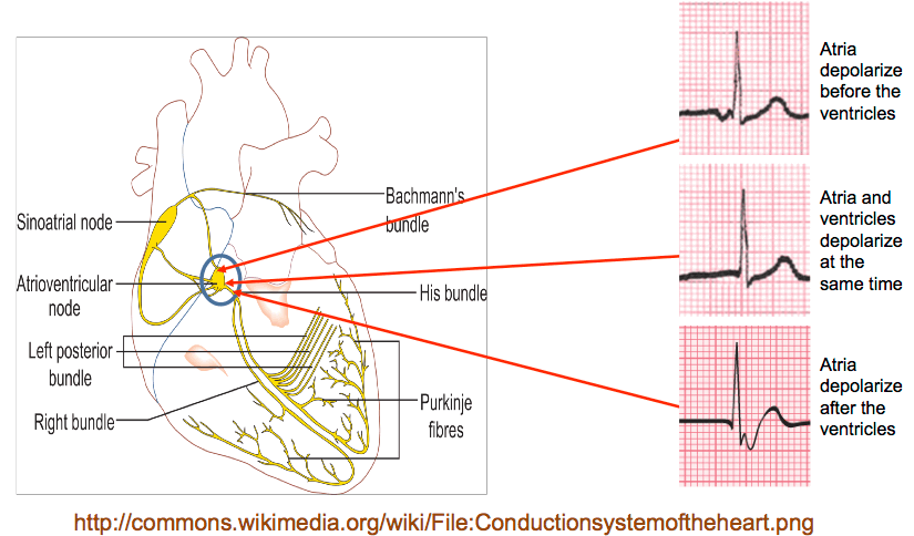
- These junctional dysrhythmias primarily affect the P wave.
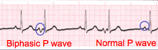
- Electrical impulses formation and flow in a normal heart follow a forward or antegrade flow through the atria. This results in the upright P wave we see with Sinus rhythms.
- With junctional rhythms, the impulse is initiated in the AV junction. This impulse point results in a backward or retrograde flow of electricity during atrial depolarization.
- This change in the flow of electricity results in an inverted P wave. This morphologic feature is unique to Junctional complexes and rhythms.
Inverted P Wave
- The result of this retrograde depolarization is the classic inverted P wave.
- Because the electrical impulse causing atrial depolarization may come from anywhere in the AV junction. This can affect the location or coordination with the QRS complex.
Lesson #4: Introduction Part 2
P Wave & QRS Complex
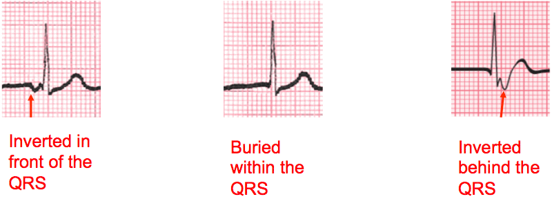
- The coordination of the P wave and QRS complex is also affected by the increased conduction velocity of the electrical impulses in the ventricular myocardium.
- The inverted P wave may be seen before or after the QRS complex, or it may be buried within it.
- Refer to the image on the next slide to reinforce this concept.
Illustration
Intro to PJC
- PJC's can occur for a number of different reasons i.e., diet, fatigue, stress, disease, ischemia to name a few.
- Premature complexes frequently occur in bradycardic rhythms, but may occur almost any time.
- PJC's occur when an early electrical impulse occurs from a location in the atria other than the SA node.
Types
We will be discussing the following complexes and rhythms:
- Premature Junctional Complexes and Junctional
- Escape Beats
- Junctional Rhythm
- Accelerated Junctional Rhythm
- Junctional Tachycardia
- Although not specifically a Junctional rhythm, we include supraventricular tachycardia because it may originate above the ventricles (including the junction).
Lesson #5: Premature Junctional Complex (PJC) and Junctional Escape Beats
Part I
- PJC’s and Junctional Escape Beats may occur for a number of different reasons i.e., diet, fatigue, stress, disease, ischemia
- Premature complexes and Junctional Escape Beats frequently occur in bradycardic rhythms, but may occur almost any time.
- Morphologically there is no difference between PJC’s and Escape Beats.
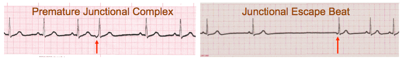
Part II
- After reviewing the previous slide you should note that the difference is that PJC’s occur early and disrupt the underlying rhythm.
- Junctional escape beats typically occur when a cardiac rhythm is too slow and a backup pacemaker site initiates an electrical impulse.
- It is fairly common that a Junctional Escape Beat will occur as the first complex to terminate Sinus Arrest
PJC Rhythm ECGs
- This early impulse causes an early cardiac complex which disrupts the underlying rhythm.
- The locus of stimulation being different, results in a change in the morphology of the P wave (inverted if it can be seen).
- PJC ECG’s can occur occasionally or frequently.
- PJC’s can be observed with or without a pattern
- The P wave with PJC’s may occur before or after the QRS complex or be buried within the QRS.
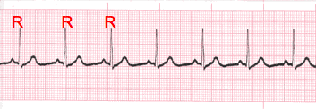
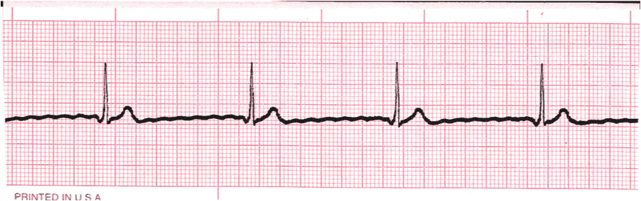
Lesson #6: PJC Tracings
Example
- The R to R interval is irregular
- The fourth complex is early
- The P wave on the early complex is inverted.
Practice Strip
Analyze this tracing using the five steps of rhythm analysis.
Show Answer
- Rhythm: Irregular
- Rate: 70
- P Wave: upright, early complex inverted
- PR interval: 0.16 sec, early complex with shorter PR
- QRS: 0.08 sec
- Interpretation: Sinus Rhythm with PJC
Lesson #7: Junctional Escape Beat
Practice Strip
>Analyze this tracing using the five steps of rhythm analysis.
Show Answer
- Rhythm: Irregular
- Rate: 40
- P Wave: upright, third complex inverted
- PR interval: 0.16 sec, third complex with shorter PR
- QRS: 0.08 sec
- Interpretation: Sinus Arrest with Junctional Escape Complex
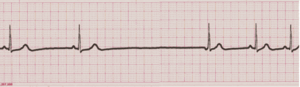
Lesson #8: Junctional Rhythm
Junctional Rhythm
- Rhythms are often named according to the origin of the electrical activity in the heart or the structure where the problem is occurring.
- Junctional Rhythm aka Junctional “Escape” Rhythm is aptly named due to the electrical impulses causing the atrial activity are originating in the AV Junction.
- The rate for this rhythm is the same as the AV Junction, 40 – 60 bpm.
- The change in the locus of stimulation affects the morphology (inverted) and potential location of the P waves (before, during or after the QRS complex).
- If the P wave occurs before the QRS, the PR interval will likely measure shorter than normal (0.12 second).
- If the P wave is buried or occurs after the QRS, it cannot be measured.
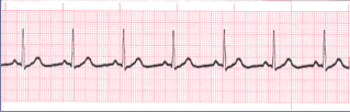
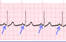
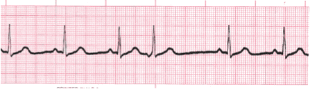
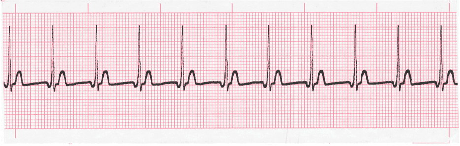
Practice Strip
Analyze this tracing using the five steps of rhythm analysis.
Show Answer
- Rhythm: Regular
- Rate: 43
- P wave: inverted
- PR interval: 0.08 sec
- QRS: 0.08 sec
- Interpretation: Junctional Escape Rhythm
Lesson #9: Accelerated Junctional Rhythm
Description
- Accelerated junctional rhythm is just a faster version of junctional escape rhythm. The criteria is the same with the only difference being the heart rate range is 60 – 100 bpm.
- Note the heart rate range. It is the same as the range for normal sinus rhythm.
- When considering the heart is a pump and if everything else is functioning properly, we should expect a patient with accelerated junctional rhythm to display signs of normal cardiac output i.e. alert and oriented, normotensive, without apparent signs of distress.
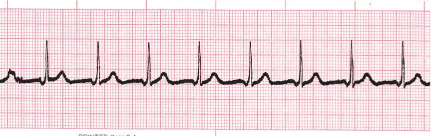
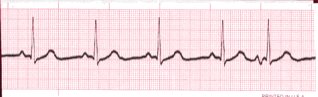
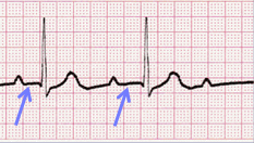
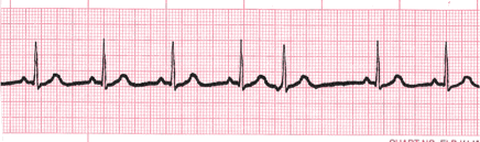
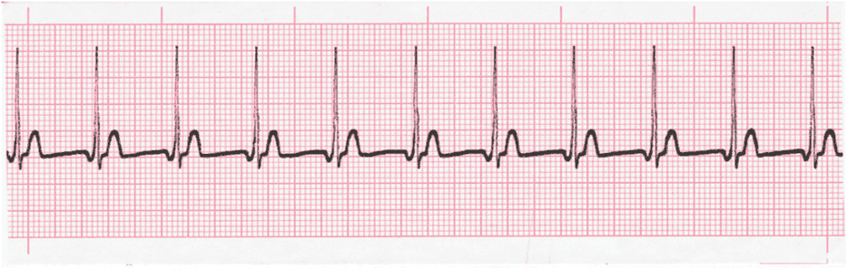
Practice Strip
Analyze this tracing using the five steps of rhythm analysis.
Show Answer
- Rhythm: Regular
- Rate: 83
- P wave: inverted
- PR interval: 0.08 sec
- QRS: 0.08 sec
- Interpretation: Accelerated Junctional Rhythm
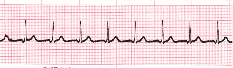
Lesson #10: Junctional Tachycardia
Junctional Tachycardia
- Junctional tachycardia occurs when a junctional rhythm exceeds 100 bpm.
- Junctional tachycardia commonly ranges from 100 – 180 bpm.
- All other criteria is the same.
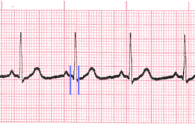
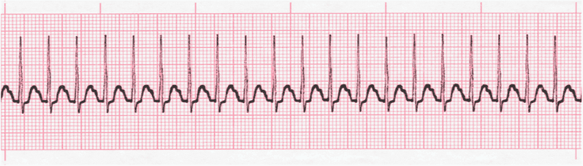
Practice Strip
Analyze this tracing using the five steps of rhythm analysis.
Show Answer
- Rhythm: Regular
- Rate: 103
- P wave: inverted
- PR interval: 0.10 sec
- QRS: 0.08 sec
- Interpretation: Junctional Tachycardia
Lesson #11: Supraventricular Tachycardia
Supraventricular Tachycardia
- Supraventricular tachycardia (SVT) occurs when the heart rhythm occurs as a result of electrical impulses initiated above the ventricles at a rate of 150 - 250 bpm or more.
- What will remain will be a normal appearing and measuring QRS complex depolarizing at a rapid rate.
- As a result of the rapid rate, the SVT ECG complexes will be very close to one another.
- The T waves of the previous SVT rhythm complex will cover the P wave of the next complex. P waves will be entirely buried making it impossible to describe their morphology and measure the PR interval.
SVT Practice Strip
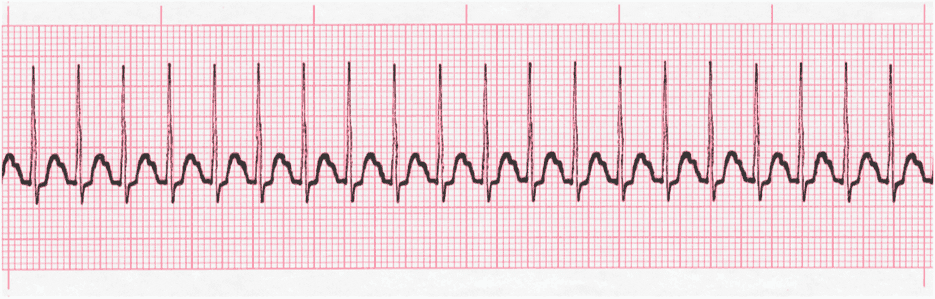
Analyze this tracing using the five steps of rhythm analysis.
Show Answer
- Rhythm: Regular
- Rate: 200
- P wave: Buried
- PR interval: Unable to measure
- QRS: 0.06 sec
- Interpretation: Supraventricular Tachycardia
Lesson #12: Quiz Test Questions 314
Question #1
When analyzing a rhythm strip, it qualifies as being regular when
B. the PR interval measures the same
C. the QRS complexes measures the same
D. the R - R intervals measure the same
Question #2
When analyzing a rhythm strip, it qualifies as being regular when
B. Rhythm regularity
C. QT Interval
D. QRS complex measurement
Question #3
Which of the following is considered normal range of the QRS complex?
B. 0.06 - 0.10 minutes
C. 0.12 - 0.20 seconds
D. 0.06 - 0.10 seconds
Question #4
>Which of the following is considered normal range of the QRS complex?
B. 0.06 - 0.10 minutes
C. 0.12 - 0.20 minutes
D. 0.06 - 0.10 seconds
Training Resources
Junctional Rhythm EKG Training
A good starting point for learning is our junctional rhythms module. This module focuses on the morphologic features and qualifying criteria of junctional rhythms. A concise summary of a five-step EKG analysis methodology is also included.
EKG Rhythm Tests
Hundreds of heart rhythms. Tests can be tailored for specific learning needs.
EKG Rhythm TestsExternal References
Authors and Reviewers
- EKG heart rhythm modules: Thomas O'Brien
-
Medical review: Dr. Jonathan Keroes, MD
- Medical review: Dr. Pedro Azevedo, MD, Cardiology
-
Last Update: 11/8/2021
Sources
-
Electrocardiography for Healthcare Professionals, 5th Edition
Kathryn Booth and Thomas O'Brien
ISBN10: 1260064778, ISBN13: 9781260064773
McGraw Hill, 2019 -
Rapid Interpretation of EKG's, Sixth Edition
Dale Dubin
Cover Publishing Company -
12 Lead EKG for Nurses: Simple Steps to Interpret Rhythms, Arrhythmias, Blocks, Hypertrophy, Infarcts, & Cardiac Drugs
Aaron Reed
Create Space Independent Publishing -
The Virtual Cardiac Patient: A Multimedia Guide to Heart Sounds, Murmurs, EKG
Jonathan Keroes, David Lieberman
Publisher: Lippincott Williams & Wilkin)
ISBN-10: 0781784425; ISBN-13: 978-0781784429
Sv: 0 | pv: 1
This website is only for professional medical education. Contact your doctor for medical care. 2026 © MedEdu LLC. All Rights Reserved. Terms & Conditions | About Us | Privacy | Email Us